
Medical
October 11, 2017
Hara Eye Hospital, Japan
Hara Eye Hospital, Utsunomiya, Japan
Ikegami MKC-704KHD 4K-output Camera Contributing to Eye Surgery
The Hara Eye Hospital is a specialised ophthalmology
The Hara Eye Hospital is a specialised ophthalmology hospital located opposite Tobu Utsunomiya railway station in Tochigi, Japan.
Cataract surgery, retinal vitreous surgery and glaucoma surgery are available, same-day and stay-over surgery can be performed, and the latest treatment can be obtained. Facilities available include diagnosis and laser-treatment of glaucoma, diabetic retinopathy, cataract removal and post-surgical inspection. Founded in 1913, Hara Eye Hospital has provided opthalmic services for over a century. It performs special diagnosis and treatment as a specialised ophthalmology hospital and also fulfils the role of being the area's 'local family doctor for eyes'. The operating room was renewed in 2017 during full renovation of the main hospital building accompanied by construction of a completely new wing. As part of this renovation, Ikegami delivered and installed a 4K surgical video system including the MKC-704KHD 4K-output camera. In addition, Ikegami installed four THD-23FHD Full-HD cameras, updating analogue camera equipment used for slit-lamp eye inspections in the consultation room. Ikegami asked Takeshi Hara MD, the fourth generation director of the Hara Eye Hospital, about the Ikegami cameras which were introduced this time.
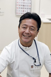
Hara Eye Hospital director Dr. Takeshi Hara.
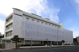
The newly renovated Hara Eye Hospital building.
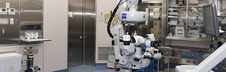
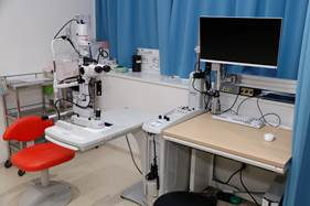
Surgical microscope and imaging system at Hara Eye Hospital.
Slit-lamp in examination room
Ikegami THD-23FHD attached to the slit-lamp
Introducing a 4K-output camera in anticipation of the future
The Hara Eye Hospital management were seeking a future-proof high-specification imaging system for a new operating room. They were introduced to the Ikegami MKC-704KHD 4K-output camera which is designed for use with surgical microscopes: "I usually see a difference in image quality between standard-definition and high-definition sources because I am viewing in HD," Dr. Hara comments. "We decided to introduce the latest video system to ensure the highest possible visual consistency. The hospital evaluated an MKC-704KHD with a 4K output, high sensitivity and four times higher resolution than a Full-HD camera. We found it both useful for ophthalmic surgery and future-proof."
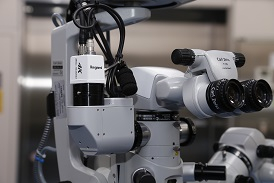
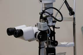
Camera head of MKC-704 KHD attached to the microscope
Patient's family is able to watch surgical process
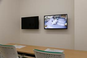
Observation room:
Families can watch surgery
during their relative's operation.
"From 1975 onward the observation room has been equipped with facilities that allow viewing of images from the operating microscope and of the operating room itself." Dr Hara adds. "In glaucoma surgery, we switch to localised lighting during surgery and reduce the room lighting. The reduced-illumination areas remain visible because the MKC-704KHD is highly sensitive. For families in the visiting room watching surgical images, our explanation has changed from 'The lights will go off' to 'The screen gets a bit darker'. The high sensitivity of MKC-704KHD enables the family watching with the patient to feel relaxed, involved and informed."
Representation of light and dark gradation indispensable for recording eye surgery
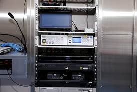
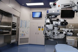
MKC-704KHD CCU and MDR-600HD recorder
installed on the surgery wall.
"The centre of the human eye is black but the surrounding area is much lighter. In cataract surgery, we concentrate on the black part of the eye. In glaucoma and conjunctivitis surgery, we also study the white area outside the pupil, also. If we try to observe the dark black part of the eye, the white parts around it will be visible unless we adjust the image precisely so that the red blood vessels of the conjunctiva do not appear. It is a very difficult image area to record. For best results, this clinic uses two modes of cataract surgery and glaucoma. Patterns appropriate for each operation are set up and switching can be done instantaneously which is very useful. We are fully accustomed to working with the Ikegami MKC-704KHD camera."
Moving the camera image closer to the photoreceptor
Hara Eye Hospital was keen that the resolution should be as high as possible but the density of human visual cells is still higher than the camera resolution. Ikegami is eager to see video technology continue advancing in line with further progress in medical treatment. In addition, we can superimpose each patient’s initials and dates on the images which will make it easier to isolate specifically desired data from hospital archives. We hope to focus on improving data organisation and retrieval methods to make the location of relevant information as easy as possible. Ikegami will embody the needs of everyone involved in the medical field and contribute to the development of medical care with visual technology.
Doctor Hara’s medical career:
1989 Graduated from Iwate Medical University.
1991 Ophthalmologist, Department of Medicine,University of Tokyo,Assistant Professor, Faculty of Education.
1993 Worked at the Omiya Red Cross Hospital University of Tokyo,Ophthalmology Glaucoma specialised outpatient clinic.
1998 Received a doctorate in medicine at the University of Tokyo.
1999 Omiya Red Cross Hospital Deputy Director of Ophthalmology.
2001 Jichi Medical University Ophthalmology course lecturer.
2005 Part-time Lecturer, Jichi Medical University.Deputy Director of Hara
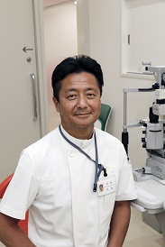
Director, Takeshi Hara